Random Local Flap – Advancement
Skin Flaps
Repair of a defect by side-to-side closure is frequently possible allowing scars to be planned in favourable skin lines.
Such a repair often requires undermining but it is essential that there is no distortion of surrounding free boundaries. When a defect cannot be closed this way, a full thickness skin graft or a skin flap may be used. Simple flaps use the skin adjacent to the defect, which is an excellent match for colour and texture. The types of flaps commonly used in dermatological surgery are random pattern, and are described according to their design and movement. There are four types commonly used, advancement, rotation, transposition and subcutaneous island pedicle flaps. There may be features of advancement and rotation in a transposition flap and advancement as well as rotation in a rotation flap. In any given site, the flap selected will depend upon local factors such as skin laxity and mobility of the adjacent skin and the texture of the surrounding skin, and the proximity of the defect to key anatomical boundaries or free margins such as the lips, eyelids and ala rim.
The surgical principles in carrying out a flap repair are common to basic wound closure – undermining, haemostasis, meticulous care in handling the skin, use of subcuticular stitches, stitching and approximation of sides of unequal height and unequal length, and the employment of dog-ear correction.
As a general rule when closing flaps the primary defect is closed first with advancement and rotation flaps, while with transposition flaps including bilobed flaps the secondary defect is closed first.
The flaps discussed here are:-
- Advancement
- Rotation
- Transposition
- Subcutaneous pedicle
Advancement Flaps
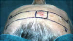
In a simple advancement flap, the movement is entirely in one direction and the flap advances over the defect. The limbs of the flap are cut to a length to breadth ratio not exceeding 3:1 on the face, or 2:1 or 1:1 ratio on the trunk.
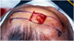
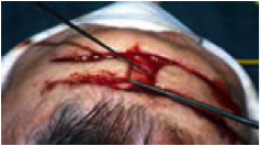
The flap consists of skin and fat and is fully undermined along with the area around the pedicle. It is advanced over the defect in a trial movement. Often a pucker (dog-ear) is seen at the base of the pedicle. These can be cut out (Burow’s triangles). Often the size of these triangles will be of different sizes on either side of the flap depending on local tissue laxity.
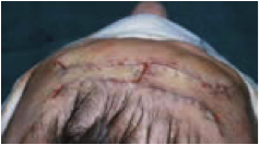
Key stitches with subcutaneous absorbable sutures secure the leading edge of the flap to the opposite side of the defect. Larger defects may require bilateral advancement. The flaps do not have to be of the same length. They may not join at the centre of the defect because of variability of the elasticity and movability of the skin.
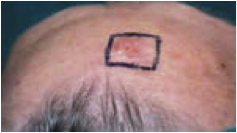
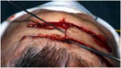
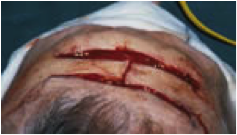
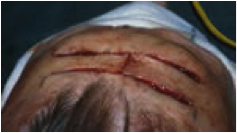
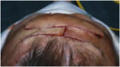
These flaps are useful on the forehead.
 |
 |
 |
 |
 |
 |
 |
 |
 |
Variations of the simple advancement flap are the A – T and the O – T and the Burow’s wedge design (see diagrams).
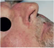
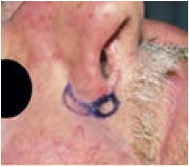
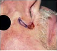
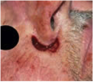
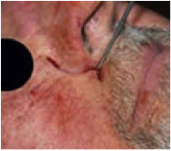
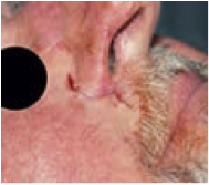
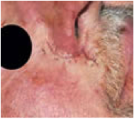
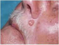
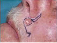
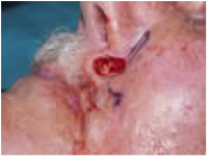
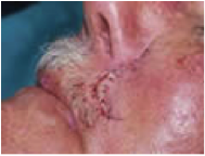
Base of nose
 |
 |
 |
 |
 |
 |
 |
|
 |
 |
 |
 |
