Fusiform Elliptical Excision
Using the Scalpel

The Pencil GripThis is useful for short, fine incisions, but because of the angle to the skin there is diminished cutting edge contact, which decreases depth and direction control in long incisions. |

The Fingertip GripThis gives less depth variation and greater direction control with long incisions; however, there is diminished fine control. |
Incision Methods

Press CuttingPress vertically downwards on stationary knife until it cuts through the tissue. This gives good control in length and direction, e.g. draining an abscess, beginning an excision biopsy. However there is poor depth control. |

Slide CuttingBlade motion at right angles to scalpel pressure. There is accuracy of depth control and precise direction and length control. However, the beginning and end of the cut tends to be shallow. |

Stabilising the SkinA stretched undistorted surface prevents jagged cuts. |
Fusiform Elliptical Excision of Lesion
• Mark clinical edge of lesion with marking pen.
• Determine the most appropriate direction for the excision using relaxed skin tension lines.
•Mark the shape of the ellipse with a length to width ratio 3:1 or greater. Ensure the clearance is adequate; measure the clearance and record it in the patient’s notes. Determine the most appropriate direction for the excisi

on using relaxed skin tension lines.
•Obtain anaesthesia and wait for the adrenaline effect.
•Stabilise the skin with the fingers and thumb.
• When starting the wound, hold the scalpel upright, so that the point is vertical as it enters the skin at one of the apices of the ellipse, and cut through the skin almost to the other apex and then repeat on the other side of the wound and finally reverse the scalpel to cut through the points at the opposite apex.
• When cutting, cut boldly along the marked line, in a single movement avoid making numerous small cuts, which will cause a jagged margin.

• Ensure scalpel is vertical to skin at start and finish. Avoid under cutting.
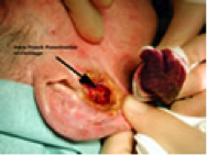
• Dissect the specimen with scissors, using a skin hook where possible to lift the specimen out of the skin. This will avoid crushing the specimen with forceps. Cut the base parallel with the skin. The shape of the specimen, and also the defect should be U-shaped and not V-shaped (see diagram).
Biopsies at Special Sites
Dermatologists are often called upon to make diagnoses of diseases which affect mucous membranes, scalp, nails, etc. It is important to obtain diagnostically useful tissue and modification of the normal biopsy technique may be needed. Here we will look at special sites:-
• Scalp and eyebrow
• Genitalia
• Eyelid
• Lip and oral mucosa
• Pinna of ear
• Nails
Scalp
Depth is important. If the incision does not go down to the galea it may not contain hair follicles and in addition closure will be more difficult. Angling the incision in the same direction as the hairs emerge from the scalp should make it possible to visualise the entire hair shafts and follicles in the histology specimen. Haemostasis can be a problem, especially if an artery in the wound edge retracts and occasionally it is necessary to strangulate such arteries by undersewing through the epidermis, under the artery and up again through the epidermis, tying the knot on the surface. Vicryl rapide 2/0 or 3/0 is useful for closing scalp biopsies because even if the knots get tangled in the hair the whole thing tends to wash off about 10 days later.
Genitalia
On the penile shaft the scalpel often drags on thin, elastic skin. Holding a piece of skin in blunt forceps allows you to snip the skin just below where it is held between the forceps producing a good biopsy specimen. Vicryl rapide is a good suture, being soft and self-absorbent. A common mistake, taking glans biopsies, is to end up with a small and traumatised biopsy. An easy method is to make 2 parallel incisions, 1cm long and 2-3mm apart, making no attempt to make the lines meet. With a pair of sharp iris scissors cut out the ribbon of tissue and put in a couple of vicryl rapide sutures. It always heals well. The same approach can be used on vulval skin.
Eyelid
Thin eyelid skin is suited to snip biopsies as described for the penile shaft.
Lip and Oral Mucosa
Try to avoid cutting across the vermilion. Otherwise, lip and oral mucosa heal very well. A chalazion clamp will prevent bleeding.
Pinna of ear
Operations on the pinna require care, skill and good haemostasis. This is not the place to describe wedge resections or helical advancement flaps. However post operative defects of the skin away from the margin can be dealt with by secondary intention healing by fenestrating the cartilage with a 4mm punch biopsy tool to allow granulating tissue to grow through from the opposite side.
 |
 |