Planning the Operative Proceedure, Basic Anatomy
Relaxed Skin Tension Lines (RSTL)
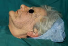 Try to place the line of the sutured wound in the relaxed skin tension lines or wrinkle lines. To find these, if they are not obvious, ask the patient to screw up their eyes, if the lesion is around the eye, or smile if the lesion is on the lower part of the face. Limbs can be bent and stretched to examine the wrinkling of the skin. If none of this manoeuvring is successful in demonstrating skin creases, try pinching up a small part of the skin near or over the lesion and look at the flow of the wrinkle lines, into and out of the pinch and adjust the direction of pinch until the lines are parallel with the pinched direction.
Danger Areas
Before undertaking any form of operative intervention operators should familiarise themselves with the structures in and immediately below the skin that could be damaged. Some of the structures that are particularly vulnerable are noted below.
a) Temporal artery. Variable route across the temple. In elderly people it is often tortuous and surrounded by very little subcutaneous fat. If damaged it should be ligated with 3/0 vicryl or dexon.
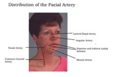
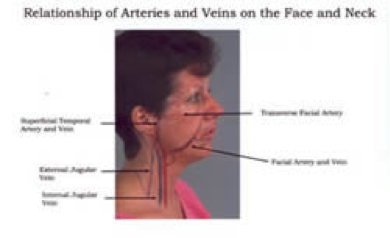
b) Facial artery. This crosses the mandible at the anterior border of the masseter muscle traverses the lower cheek to a point just lateral to the angle of the mouth and gives off the superior and inferior labial arteries. It then runs up the side of the nose to terminate around the medial canthus of the eye.
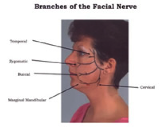
c) Facial nerve. The temporal branch frequently subdivides shortly after leaving the parotid gland. These branches are very superficial and easily damaged. The marginal mandibular branch is most at ri
sk where it crosses the inferior border of the mandible adjacent to the facial artery at the anterior border of masseter.
d) Superficial branches of the trigeminal nerve. Especially vulnerable are the supratrochlear and supraorbital nerves, the zygomaticotemporal nerve and the auriculotemporal nerve on the lateral cheek. In the lower part of the face the mental branch is vulnerable on the chin after it leaves the mental foramen.
e) Spinal Accessory Nerve. Runs across the base of the posterior triangle emerging from the posterior border of sternocleidomastoid at the junction of upper and middle thirds. It courses posteriorly inferiorly and disappears behind the trapezius muscle in the lower third of the triangle. It is deep to superficial fascia and hence is less likely to be damaged than some of the nerves of the face that lie more superficially.
Upper Limb
Care should be taken in the axilla where nerves arising from parts of the brachial plexus are near the surface. At the wrist the radial artery and many other important structures are present superficially and the operator should refer to an anatomical text to become acquainted with possible dangers.
Lower Limb
The femoral triangle contains nerve artery and vein all of which are superficial. The lateral cutaneous nerve of the thigh is easily damaged over the upper outer quadrant.
At the knee the lateral popliteal nerve is palpable and superficial where it rounds the head of the fibula. However the popliteal fossa is also an area to be approached with caution. In the lower leg perhaps the greatest risk is from varicose veins that need to be tied off adequately if damaged. Around the ankle there is a little fat and hence all structures are more vulnerable.
The above is not intended to intimidate the dermatological surgeon as most of the surgical operations will be superficial and well away from any danger areas. However it is essential to know the anatomy of the area in which you are working.
Skin Marking

Having considered the relaxed skin tension lines and the danger areas, the skin should now be marked up with a skin marking pen, using Bonney’s blue as the marking medium, ordinary felt tip pens should be avoided as some contain particles which may tattoo the skin if cut through. It is frequently helpful, particularly with malignant lesions to mark around the clinical margin of the lesion and then to mark outside to establish the margin of clearance. At this stage it is also worthwhile pinching up the skin to make sure that there will be sufficient spare skin for closure of the wound.
