Currettage
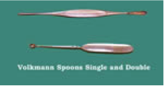
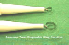
Two types of curette are used.
In the UK the spoon or Volkmann type predominates but recently the ring curette has been preferred having the advantage that excess soft tissue can escape through the ring. They are usually 3-7mm in diameter.
Very small, or “ophthalmic” spoon curettes (1-2mm) are extremely useful for small lesions and for probing small tumour extensions.

Theseinstruments must be kept sharp.
Advantages –
- Simple, cheap, quick
- Good cosmetic results with superficial lesions
- Good cure rates with small, non-fibrotic tumours
- Sutures, and sometimes anaesthetic, not required.
Disadvantages –
- Secondary intention healing may be slow.
- The scar is usually hypo pigmented.
- Poor cure rates with large, fibrotic or recurrent tumours and at certain sites e.g. ear, naso-labial.
Indications
Usually for removal of small growths on the skin surface: –
- Viral warts
- Basal cell papillomas (Seborrhoeic keratoses)
- Pyogenic granulomas
- Actinic keratoses and Bowen’s disease
- Small BCC’s and SCC’s (less than 1cm diameter)
- “Curette” biopsy
- Determination of extent of tumour before formal excision.
Contra-indications
- Fibrotic or scarring lesions, e.g. morphoeic BCC’s.
- Recurrent tumours, except as palliative treatment
- Large tumours (more than 1cm diameter)
- Tumours penetrating through the dermal sling
- Malignant melanomas
Technique
- For small lesions local anaesthetic is not necessarily required – the anaesthetic may be more painful than the curettage.
- Tension the skin with fingers to provide firm surface.
- Hold the curette like a pen.
- Scoop the lesion out with downward rotary motion.
- Scrape all the abnormal tissue away from the base and all round the periphery. It usually separates fairly easily from the normal surrounding skin, but must be done thoroughly.
- Control bleeding – usually by cautery or electrodessication
- Repeat 5 and 6 if the lesion is malignant
- If the lesion is benign use very light cautery only to obtain haemostasis, or a weak styptic such as aluminium chloride hexahydrate 20% (Anhydrol forte) applied with a cotton bud.
- Dressings are not necessarily required, but patient instruction (qv) is helpful.
- Send specimen to histopathology laboratory.
When treating malignant lesions, you should abandon the technique if the dermis is breached and a formal excision should be undertaken.
